Notes
|
Topics to be learn :
|
Terms related to spherical mirrors :
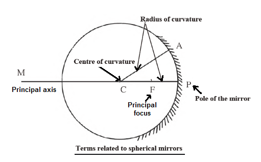
- Construction of concave mirrors : A hollow spherical glass can be transformed into a concave mirror by polishing its inner surface or coating its outer side with a thin silver layer and painting it red.
- Construction of convex mirrors : A hollow spherical glass can be transformed into a convex mirror by polishing its outer surface or coating its inner side with a thin layer of silver and painting it red.
Lenses :
Lens : A lens is a transparent material bound by two surfaces, out of which at least one surface is spherical.
Difference between lens and mirror :
- A mirror has one reflecting surface. By reflection of light, it forms an image of the object placed in front of it. A mirror is not transparent.
- A lens has two surfaces that form an image by refraction of light. A lens is transparent.
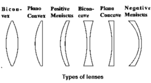
Types of lenses : Different types of lenses are shown in figure.
Terms of lenses :
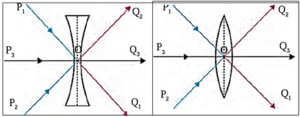
- Centre of curvature (C) of a lens : The centres of the spheres whose parts form the surfaces of a lens are called the centres of curvature of the lens. A lens has two centres of curvature C1 and C2 for its two spherical surfaces.
- Radii of curvature (R1, R2.) of a lens: The radii of the spheres whose parts form surfaces of a lens are called the radii of curvature of the lens.
- Principal axis of a lens: The imaginary straight line passing through the two centres of curvature of a lens is called the principal axis of the lens. |
- Optical centre (O) of a lens : The point inside a lens on the principal axis, through which light rays pass without changing their path is called the optical centre (O) of the lens.
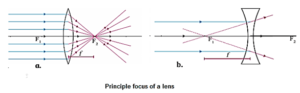
- Principal focus (F) of a lens : When light rays parallel to the principal axis are incident on a convex lens, they converge at a point on the principal axis. This point is called the principal focus (F) of the convex lens. Light rays travelling parallel to the principal axis of a concave lens diverge after refraction in such a way that they appear to be coming out of a point on the principal axis. This point is called the principal focus of the concave lens. A lens has two principal foci F1, and F2.
- Focal length (f) of a lens: The distance between the optical centre and the principal focus of a lens is called the focal length (/) of the lens.
Ray diagram for refracted light :
Ray diagrams help in obtaining images formed by lenses, including their position, size, and nature.
Convex lens:
- A lens having both spherical surfaces puffed up outwards is called a convex lens or double convex lens or biconvex lens.
- A lens having one surface plane and the other (spherical surface) bulging outward is called a planoconvex lens.
- A convex lens is thicker in the middle than at the edges.
- It is a converging lens.
- The concavo-convex lens has one spherical surface concave and the other convex such that it behaves as a convex lens.
Rules for obtaining an image formed by a convex lens :
Rules for obtaining an image formed by a convex lens :
(i) When the incident ray is parallel to the principal axis, the refracted ray passes through the principal focus.
(ii) When the incident ray passes through the principal focus, the refracted ray is parallel to the principal axis.
(iii) When the incident ray (ray of light) passes through the optical centre of the lens, it passes without changing its direction.
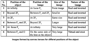
Images formed by convex lenses :
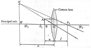
(i) Object at infinity : In this case, the image is formed at focus F2 of the convex lens. It is real, inverted and highly diminished (point-sized).
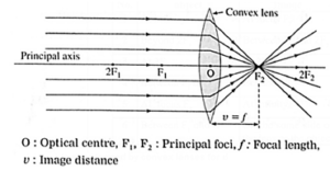
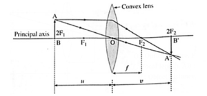
(ii) Object beyond 2F1 : In this case, the image is formed between F2 and 2F2. It is real, inverted and diminished.
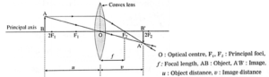
(iii) Object at 2F1 : In this case, the image is formed at 2F2. It is real inverted and of the same size as that of the object.
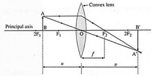
(iv) Object between F1 and 2F1 : In this case, the image is formed beyond 2F2. It is real, inverted and magnified (enlarged).
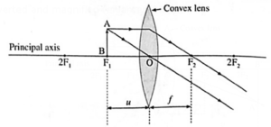
(v) Object at focus F1 : In this case, the image is formed at infinity. It is real, inverted and infinitely large (highly magnified).
(vi) Object between focus F1 and optical centre O : In this case, the image is formed on the same side of the lens as the object. It is virtual, erect and larger than the object.
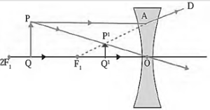
Images formed by concave lenses :
- If the object is at infinity, the image is formed at the focus of the lens, on the same side of the lens as the object. It is virtual, erect and much smaller than the object (point image).
- If the object is at any finite distance from the lens, the image is formed on the same side of the lens as the object and between the focus and the optical centre of the lens. It is virtual, erect and smaller than the object. The image distance is less than the object distance.
Rules used for drawing ray diagrams for the formation of an image by a concave lens :
- When the incident ray is parallel to the principal axis, the refracted ray, when extended backwards, passes through the principal focus.
- When the incident ray is directed towards the principal focus F2, the refracted ray is parallel to the principal axis.
- When the incident ray passes through the optical centre of the lens, it passes without changing its direction.
Characteristics of an image formed by a concave lens :
The image formed by a concave lens is always virtual, erect and smaller than the object. It is on the same side of the lens as the object.
Generally, it is formed between the optical centre of the lens and the principal focus F1 If the object is at infinity, the image is a point image formed at F1.
Sign convention :
In this case, the optical centre (0) of the lens is taken as the origin and the principal axis of the lens is taken as X-axis of the coordinate system.
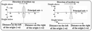
- The object is always placed at the left of the lens. All distances parallel to the principal axis are measured from the optical centre of the lens.
- All distances measured to the right of the origin are taken as positive while distances measured to the left of the origin are taken as negative.
- Distances measured perpendicular to and above the principal axis are taken as positive.
- Distances measured perpendicular to and below the principal axis are taken as negative.
- The focal length of a convex lens is positive and that of a concave lens is negative. (Fig.)
Lens formula:
The relationship between the object distance (u), image distance (v) and focal length (f) of a lens is called the lens formula and is given as
\(\frac 1f = \frac 1v - \frac 1u\)
(i) Magnification by a lens:
The magnification (M) produced by a lens = \(\frac{\text{height of the image (h_2)}}{\text{height of the object (h_1)}} = \frac vu \)
- Magnification is positive for a virtual image and negative for a real image.
(ii) Power of a lens : The power (P) of a lens = \(\frac{1}{\text{focal legth (f) of the lense}}\)
- If the powers of the two lenses are P1 and P2 then the effective power of their combination is P = P1 + P2. Thus, when two lenses are kept touching each other, the power of the combined lens is equal to the sum of their individual powers.
- Its SI unit is the dioptre (D). If f = 1 metre, P = 1 dioptre.
(iii) Combination of lenses: If two lenses with focal lengths f1 and f2 are kept in contact with each other, the effective focal length of the combination, f, is given by
\(\frac 1f = \frac{1}{f_1} + \frac{1}{f_2}\)
The effective power of the combination of the lenses, P, is given by P = P1 + P2, where P = \(\frac{1}{f}\), P1 = \(\frac{1}{f_1}\), P2 = \(\frac{1}{f_2}\),
Working of human eye and lens :
Shape and the size of the human eyeball : The human eyeball is approximately spherical in shape with a diameter of about 2.4cm.
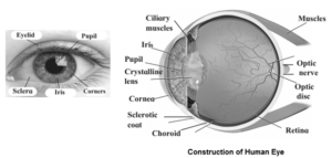
Retina : The retina is a light-sensitive screen with a delicate membrane and numerous light-sensitive cells that provide information about an object's brightness and color. When light hits the retina, these cells activate and generate electrical signals, which are then passed to the brain via optic nerves. The brain interprets these signals, allowing us to perceive the object as it is.
Ciliary muscles : The muscles which hold the eye lens in its position, and bring about changes in the shape (curvature) of the eye lens, and hence of focal length are known as ciliary muscles.
Cornea: The cornea is a thin and transparent cover (membrane) on the human eye through which light enters the eye. Maximum refraction of light rays entering the eye occurs at the cornea.
Iris : The iris is a dark fleshy screen (muscular diaphragm) behind the cornea in the human eye. Its colours are different for different people.
Pupil : The pupil, a circular opening at the iris's center, controls and regulates light entering the eye. It contracts when too much light is present and dilates when insufficient, adjusting the amount of light entering the eye.
- Adaptation : The tendency of the pupil in the human eye to adjust the opening for light, depending on the intensity of incident light, to control and regulate the amount of light entering the eye is called adaptation.
Eye Lens : A transparent biconvex crystalline lens is located just behind the pupil in the human eye. The eye lens provides small adjustment of focal length to form a real and inverted sharp image on the retina.
Persistence of vision : The image of an object remains imprinted on our retina for =1/16th of a second after the object is removed from the sight. The sensation on the retina persists for a while. This is called persistence of vision.
Power of accommodation of the eye: The ability of the eye lens to adjust its focal length is called the power of accommodation of the eye.
The minimum distance of distinct vision and the near point : The minimum distance from the normal eye at which an object is clearly visible without stress on the eye is called the minimum distance of distinct vision. It is 25 cm for the normal human eye.
Defects of vision and their correction :
Problems of vision are related to (i) weakening of ciliary muscles (ii) change in the size of the eyeball (iii) irregularities on the surface of cornea (iv) formation of a membrane over the eye lens.
(i) Myopia or Nearsightedness :
Myopia is a vision defect where the human eye can see nearby objects but not distant ones clearly. This results in an image of a distant object formed in front of the retina. Correction : To correct myopia, a concave lens is used to diverge light rays before they hit the eye lens, forming the image on the retina after the lens's action.
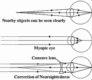
Possible reasons of myopia :
- The curvature of the cornea and the eye lens increases. The muslces near the lens cannot relax so that the converging power of the lens remains large.
- The distance between the eye lens and the retina increases as the eyeball elongates.
(ii) Hypermetropia or Farsightedness : Hypermetropia, or farsightedness, is a vision defect where the human eye can see distant objects but not nearby ones. This results in the image of a nearby object falling behind the retina.
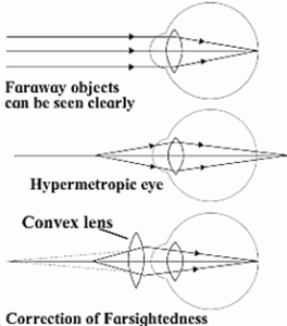
Correction : Hypermetropia is corrected using a sultable convex lens, which converges light rays before striking the eye lens. The correct focal length is chosen to achieve the required convergence, forming the image on the retina.
Possible reasons of hypermetropia:
- Curvature of the cornea and the eye lens decreases. Hence, the converging power of the eye lens becomes less.
- The distance between the eyelens and retina decreases (relative to the normal
- eye) and the focal length of the eye lens becomes very large due to the flattening of the eyeball.
Presbyopia (also called old age hypermetropia) : Presbyopia is a vision defect where elderly individuals struggle to see nearby objects without glasses.
Correction : It is corrected using a convex lens that converges light rays before they fall on the eye lens, forming the image on the retina.
Reason of presbyopia : The power of accommodation of eye usually decreases with ageing. The muscles near the eye lens lose their ability to change the focal length of the lens. Therefore, the near point of the eye lens shifts farther from the eye.
Bifocal lens :
- A bifocal lens is a lens of which the upper part is a concave lens to correct myopia and the lower part is a convex lens to correct hypermetropia.
- A person suffering from myopia as well as hypermetropia, uses a bifocal lens. Nowadays, the defects of vision such as myopia and hypermetropia can be corrected using contact lenses or by laser surgery.
Camera and Human Eye :
Camera and Human Eye :
Similarities :
- In the case of a camera as well as the human eye, it is possible to control the amount of incoming light with the help of a diaphragm and an aperture.
- Both use a convex lens for focusing.
- The photographic film in a camera is coated with a photosensitive material. The retina in the eye consists of a large number of light sensitive cells.
- The photographic film in a camera is processed using chemicals and then prints (photographs) can be obtained using the appropriate paper.
- In the human eye, the electrical signals generated by light sensitive cells are passed by optic nerves to the brain which interprets them.
Differences :
- Cameras come in a variety of sizes and shapes unlike the human eye.
- Unlike the human eye, a wide variation in exposure time is possible in the case of cameras.
- The human eye is sensitive in the visible region (red to violet) of the electromagnetic spectrum, while a much wider range of the electromagnetic spectrum can be covered with cameras designed for specific purposes.
- In comparison with the human eye, a wider view and range can be covered by a camera.
- In comparison with the human eye, a wider intensity (of light) range can be covered with a camera.
- The retina is indispensable in the human eye, while cameras without a photographic film have been designed with the help of photosensitive materials and are in current use.
- With advances in technology, improved cameras are designed all the time, and the list of differences between the human eye and a camera in general would be practically endless.
Apparent size of an object :
- An object appears small or big depending upon the size of its image formed on the retina of the eye.
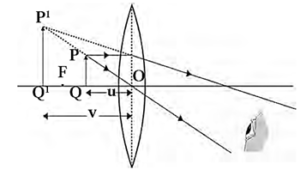
- The size of an object as perceived by the eye is called the apparent size of the object. Consider two objects of the same size, one held near the eye and the other away from the eye as shown in the following figure (Fig.). The nearby object (PQ) appears larger than the distant object (P1 Q1).
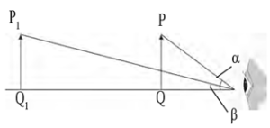
- Also, the angle ∝ subtended by the nearby object at the eye is larger than the angle β subtended by the distant object at the eye. This shows that the apparent size of an object at the eye is determined by the angle subtended by the object at the eye.
- The closer the object is to the eye, the larger its apparent size is. Conversely, the smaller the angle subtended by the object at the eye, the smaller its apparent size is.
Uses of lenses :
Use of concave lenses :
- Medical equipments, scanner, CD player – These instruments use laser light. For proper working of these equipment concave lenses are used.
- The peep hole in door- This is a small safety device which helps us see a large area outside the door. This uses one or more concave lenses.
- Spectacles- Concave lenses are used in spectacles to correct nearsightedness.
- Torch- Concave lens is used to spread widely the light produced by a small bulb inside a torch.
- Camera, telescope and microscope- These instruments mainly use convex lenses. To get good quality images a concave lens is used in front of the eyepiece or inside it.
Use of convex lenses :
Simple microscope : A convex lens with small focal length produces a virtual, erect and bigger image of an object as shown in the figure. Such a lens is called simple microscope or magnifying lens.
- The object is placed in front of the convex lens of short focal length such that the object distance is less than the focal length. The image is virtual and larger than the object. It is formed on the same side of the lens as the object.
- One can get a 20 times larger image of an object using such microscopes.
- These are used for watch repair, testing precious gems and finding their defects. A simple microscope (called a magnifying glass) is also used to read words in small print.
Compound microscope :
- A compound microscope is made of two convex lenses: objective and eye piece. The objective has smaller cross-section and smaller focal The eye piece has bigger cross-section, its focal length is also larger than that of the objective. The lenses are fitted inside a metallic tube in such a way that the distance between can be changed.
- Higher magnification can be obtained by the combined effect of the two lenses.
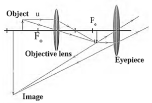
Working :
- The object is illuminated and placed in front of the objective lens, slightly beyond its focal length. The objective lens forms a real, inverted, and enlarged image on the other side.
- This intermediate image lies within the eyepiece's focal length and serves as an object for the eyepiece. The final image is virtual, highly enlarged, and inverted, formed at the minimum distance of distinct vision from the eyepiece. The final image is observed by keeping the eyepiece close.
Uses of compound microscope :
- It is used to observe blood corpuscles, plant and animals cells, microorganisms like bacteria, etc.
- It is used in a pathological laboratory to observe blood, urine, etc.
- It is a part of a travelling microscope used for measurement of very small distance.
Telescope :
Telescopes are of two types.
- Refracting telescope – This uses lenses
- Reflecting telescope – This uses mirrors and also lenses.
In both of these, the image formed by the objective acts as object for the eye piece which forms the final image.
Construction of a refracting telescope :
- A refracting telescope consists of two convex lenses, the objective lens and the eyepiece, which are directed towards the object.
- The objective lens is placed on one end of a long metal tube, while the eyepiece is placed on the outer end of a smaller tube.
- The distance between the eyepiece and the objective lens can be adjusted using a screw, and the principal axes of the lens and eyepiece are along the same line.
- The telescope is typically mounted on a stand.
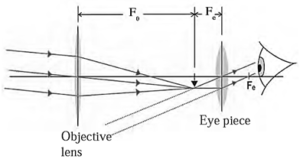
Working:
- The objective lens is used to observe distant objects by capturing the light rays from the object.
- It collects the maximum amount of light, forming a real, inverted, and diminished image in the focal plane.
- The eyepiece is adjusted to fit this image within the focal length of the eyepiece, serving as the object for the microscope.
- The final image is highly magnified, virtual, and inverted with respect to the original object.
- The final image can be observed by keeping the eye close to the eyepiece. If the image forms in the focal plane, the final image is formed at infinity.
Uses : Telescope is used to see distant objects clearly in their magnified form. The telescopes used to observe astronomical sources like the stars and the planets are called astronomical telescopes.
Optical instrument : Convex lenses are used in various other optical instruments like camera, projector, spectrograph etc.
Persistence of vision :
The eye lens creates an image on the retina, which remains on the retina as long as the object is in front of us. The image disappears when the object is removed, but this is not instantaneous and remains imprinted for 1/16th of a second, causing the sensation on the retina to persist for a while. This is called persistence of vision.
It is due to persistence of vision that we continue to see the object in its position for about 1/16th of a second after it is removed.
Examples:
- When a burning stick of incense is moved fast in a circle, a circle of red light is seen
- The working of a television set and motion picture is based on the phenomemon of persistence of vision.
- In motion pictures, photographs of moving objects are taken at the rate of more than sixteen pictures per second. These photographs are projected on the screen at the same rate. Each picture is slightly different from the other. As a result of persistence of vision, we get the impression of observing the objects in continuous motion.
How do we perceive different colours? :
How do we perceive different colours?
- The retina in our eyes contains light-sensitive cells, shaped like rods and cones.
- Rod cells respond to light intensity, providing information about object brightness or dimness.
- Conical cells respond to color, providing color information to the brain.
- The brain processes this information, resulting in the actual image of the object.
- Rod cells respond to faint light, while conical cells respond differently to red, green, and blue colors.
- Some people lack conical cells, resulting in color blindness. However, their eye sight remains normal, and they cannot distinguish between different colors.
Thnx sir